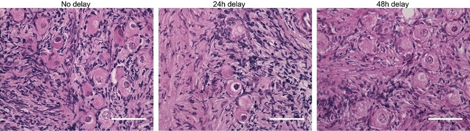
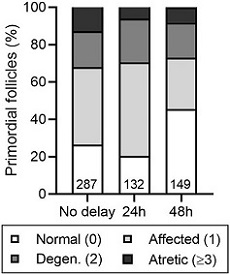
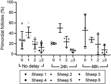
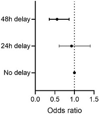
| Figure 4: Effect of delayed processing on primordial follicle morphology in cryopreserved-thawed ovarian tissue. Ovine ovarian tissue was either processed immediately (no delay; n = 6), 24 h (n = 3) or 48 h (n = 3) after collection, cryopreserved by slow freezing, thawed and fixed in Bouin’s solution. (a) Representative images of cryopreserved-thawed ovine ovarian tissue. Scale bar: 50 μm. (b) Health distribution of primordial follicles in cryopreserved-thawed ovine ovarian tissue. Numbers at the base of columns refer to the total number of follicles analysed. Primordial follicles (oocyte surrounded by a single layer of flattened pregranulosa cells) were analysed using H&E staining and scored based on the presence of morphological markers (dense chromatin, eosinophilia, shrunken ooplasm [one point each] or pyknosis [two points]) as normal (0), affected (1), degenerating (2) or atretic (≥ 3). (c) Same data as in (b), showing the distribution of health grades between animals (mean ± SD). (d) Proportional ORs and 95% CIs of follicles being graded as ≥ 1 when processed after 24 h or 48 h compared to the no delay group. Black CI bars indicate P < .05. |