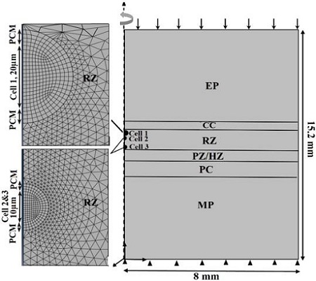
| Figure 1: The idealized axisymmetric model shows tissue and cell levels for cells positioned at three locations within the RZ. The tissue was subjected to a displacement of 15% of the cartilage thickness parallel to the axis of axisymmetry. It is restrained at the bottom (in the y-direction). It includes the epiphysis (EP), subchondral bone (SB), calcified cartilage (CC), growth plate cartilage reserve zone (RZ), and proliferative/hypertrophic zone (PZ/HZ), zone of provisional calcification (PC), and metaphysis (MP). The microscale model consists of three chondrons embedded in the extracellular matrix, ECM of the RZ. All chondrons are composed of chondrocyte cells enveloped by a pericellular matrix (PCM). GP thickness = 2 mm. |