• About the Journal |
  • Submit a Manuscript |
  • Published Articles
  • Home |
  • Journals |
  • About Us
  • Abstracting and Indexing
  • Aims and Scope
  • Article Processing Charges
  • Author Guidelines
  • Contact Information
  • Editorial Board
  • Editorial Workflow
  • Email Alerts
  • Ethics of Publication
  • Open Access
  • Open Special Issues
  • Our Policy
  • Published Articles
  • Reviewer Guidelines
  • Subscription Information
Journal of Transition Metal Complexes
Vol. 3 (2020), Article ID 236087, 9 pages
doi:10.32371/jtmc/236087
Research Article

Schiff Bases and their Copper(II) Complexes Derived from Cinnamaldehyde and Different Hydrazides: Synthesis and Antibacterial Properties

Zong Chin Khor,1,2 May Lee Low,3 and Irene Ling1,2

1School of Science, Monash University Malaysia, 47500 Bandar Sunway, Selangor, Malaysia
2Tropical Medicine and Biology Platform, Monash University Malaysia, 47500 Bandar Sunway, Selangor, Malaysia
3Department of Pharmaceutical Chemistry, School of Pharmacy, International Medical University, 57000 Kuala Lumpur, Malaysia
Address correspondence to Irene Ling, [email protected]

Received 3 December 2019; Revised 27 July 2020; Accepted 30 July 2020; Published 28 August 2020

  • Download PDF
  • Supplementary Material

How to Cite this Article

Zong Chin Khor, May Lee Low, and Irene Ling, Schiff Bases and their Copper(II) Complexes Derived from Cinnamaldehyde and Different Hydrazides: Synthesis and Antibacterial Properties, Journal of Transition Metal Complexes, 3 (2020), art236087. doi:10.32371/jtmc/236087

Abstract

New hydrazone Schiff base ligands were synthesized from the condensation of cinnamaldehyde and three different hydrazides (benzhydrazide, isoniazid, and 4-phenylsemicarbazide), and these compounds were further reacted with copper(II) ion affording three different Schiff base metal complexes. All hydrazone Schiff base ligands and their respective metal complexes were chemically characterised using NMR, FTIR, CHN analyses, and UV-visible spectrophotometry and these chemical analyses showed successful coordination of hydrazone Schiff base ligands to copper(II) ion. All synthesized Schiff base ligands and metal complexes were screened against Staphylococcus aureus, Acinetobacter baumannii, Escherichia coli, and Pseudomonas aeruginosa. Interestingly, all Schiff base metal complexes showed a higher antibacterial activity than their parent hydrazone Schiff base ligands; as shown by an increase of up to 60% of inhibition observed for one of the metal complexes.

Keywords

Schiff base; cinnamaldehyde; hydrazide; bacteria; transition metal

1. Introduction

The infection of microorganism has caused massive economic losses and threats to the human population [1]. One of the causes is the inappropriate uses of antibiotics notably mishandling that have caused a wide range of bacteria to develop antibiotic resistance [2]. Due to the fastgrowing rate of bacteria that are resistant against available antibiotic drugs, the development of a new and effective antibacterial drug is vital, as cure or therapies to common infections would be problematic in future if the issue is not resolved [2]. It is worth noting that selected antimicrobial drugs that are currently available in the market have some drawbacks such as low effectiveness, undesired side effects, and its toxicity to the human body [3]. Hence, the interest in searching for novel active compounds derived from natural products that have effective antimicrobial activities have significantly increased over the years as the antimicrobial properties of natural products often cover a wider range of bacteria with minimum side effects [4].

Schiff bases (also known as hydrazones) are extensively studied in the area of medicinal, organic, and inorganic chemistries as the C=N linkage is important in enhancing certain biological activities [5,6]. Schiff bases, typically formed from the condensation of a primary amine and an aldehyde, are deemed crucial in pharmaceutical industry owing to their effectiveness in resolving some of the biological related ailments [7]. Schiff bases can form coordinate bonds efficiently with transition metals via the lone pair electron on the azomethine nitrogen atom and oxygen atom or another heteroatom present in the Schiff base derivatives [8]. Schiff base metal complexes involving the transition metals in the first row in the periodic table, namely cobalt(II), copper(II), and nickel(II), were widely studied and their importance in improving selected pharmaceutical activities is evident [9,10,11]. To date, most of the transition metal complexes involving Schiff base ligands have proven to exhibit strong biological potencies notably in antibacterial, anti-inflammatory, antifungal, antiviral, antimalarial, antiproliferative, antitubercular, and antipyretic properties [6,12,13,14,15]. Zayed et al. concluded that Schiff base metal complexes have a greater antimicrobial effect than the unbound ligand [9]. Geeta et al. conducted antibacterial activity on gram-positive type of bacteria such as Staphylococcus aureus and gramnegative bacteria such as Escherichia coli with copper(II) and nickel(II) Schiff base metal complexes in the presence of antibiotics such as streptomycin and ampicillin [16]. The zone of inhibition for Schiff base metal complexes for both copper(II) and nickel(II) showed a stronger antibacterial activity compared to antibiotics as the zone of inhibition range for Schiff base metal complexes is much larger as opposed to when antibiotics were used.

In the present work, we have prepared and characterised three new Schiff base compounds from three different hydrazides: (i) benzhydrazide, (ii) isonicotinic acid hydrazide (isoniazid), and (iii) 4-phenylsemicarbazide, along with cinnamaldehyde. Subsequently, the Schiff base compounds were reacted with copper(II) acetate salt and each complex was tested for their bioactivity performances. Cinnamaldehyde was chosen to prepare these ligands owing to its well-known rich bioactive properties [17,18,19,20]. For example, Wang et al. studied modified cinnamaldehyde with amino acid Schiff bases and reported that these compounds showed potential in antibacterial properties (S. aureus and E. coli), and good antifungal properties (Aspergillus niger and Penicillium citrinum) where at least a 40 mm zone of inhibition (in diameter) was observed at 0.025 mol/L [21].

2. Methods

All reagents and solvents were purchased from Sigma-Aldrich and were used as received. Bacterial strains were purchased from the American Type Culture Collection.

2.1. Synthesis of Schiff bases, L1, L2, and L3

Hydrazone Schiff base ligands were prepared by directly mixing an equimolar ratio (1:1) of either benzyhydrazide, isonicotinic acid hydrazide (isoniazid) or 4-phenylsemicarbazide with cinnamaldehyde (Scheme 1) in 20 mL of hot acetonitrile and heated under reflux at 60 °C for 2 h, affording N′-((1E,2E)-3-phenylallylidene)benzohydrazide (L1), N′-((1E,2E)-3-phenylallylidene)isonicotinohydrazide (L2), and (E)-N-phenyl-2-((E)-3-phenylallylidene)hydrazidecarboxamide (L3), respectively. The hydrazone compounds formed are poorly soluble and they precipitate out as solids overnight in cold temperature (4 °C). The yellowish precipitates are easily separable through filtration and the compounds were washed with cold acetonitrile and subsequently dried at room temperature. The purity for all compounds was inspected using 1H and 13C-NMR along with melting point measurement. Attempts to crystallise the hydrazone Schiff base ligands for singlecrystal diffraction study were not successful. The chemical shifts for L1, L2, and L3 are seen as follows:

sch1
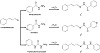
Scheme 1: Synthesis of L1, L2, and L3 from cinnamaldehyde and three different hydrazides.

L1: 1H-NMR (400 MHz, DMSO-d6) δ ppm: 11.73 (s, 1H, N-H), 8.20 (d, 1H, C-H), 7.86 (d, 2H, Ar-H), 7.60 (d, 2H, Ar-H), 7.55 (d, 1H, C-H), 7.49 (t, 2H, Ar-H), 7.37 (t, 2H, Ar-H), 7.30 (t, 1H, Ar-H), 7.04 (m, 2H, Ar-H; C-H); 13C-NMR (400 MHz, DMSO-d6) δ ppm: 163.54, 150.30, 139.60, 136.44, 133.92, 132.29, 129.39, 129.02, 128.15, 127.65, 126.20; IR: ν(cm−1) = 3,267 (m, NH), 1646 (s, C=O), 1,487 (m, C=N); Tm = 199–202 °C.

L2: 1H-NMR (400 MHz, DMSO-d6) δ ppm: 11.94 (s, 1H, N-H), 8.75 (d, 2H, Ar-H), 8.21 (d, 1H, C-H), 7.78 (d, 2H, Ar-H), 7.61 (d, 2H, Ar-H), 7.38-7.30 (m, 3H, Ar-H), 7.07 (m, 2H, C-H); 13C (400 MHz, DMSO-d6) δ ppm: 162.01, 151.52, 150.86, 140.97, 140.50, 136.31, 129.40, 127.76, 125.90, 122.07; IR: ν(cm−1) = 3,243 (m, NH), 1,651 (s, C=O), 1,491 (m, C=N); Tm = 198–201 °C.

L3: 1H-NMR (400 MHz, DMSO-d6) δ ppm: 10.66 (s, 1H, N-H), 8.76 (s, 1H, N-H), 7.75 (d, 1H, C-H), 7.60-7.51 (m, 4H, Ar-H), 7.38-7.22 (m, 6H, Ar-H), 6.95 (m, 2H, C-H, Ar-H); 13C-NMR (400 MHz, DMSO-d6) δ ppm: 153.35, 143.46, 139.63, 137.61, 136.60, 129.43, 129.08, 127.29, 125.98, 122.86, 119.81; IR: ν(cm−1) = 3,341 (m, NH), 1,670 (s, C=O), 1,445 (m, C=N); Tm = 175–177 °C.

2.2. Synthesis of Schiff base metal complexes

A 0.5 mol equivalent of copper(II) acetate monohydrate in acetonitrile was added dropwise into a solution containing 1 mol equivalent of a Schiff base compound (either L1, L2 or L3) that was dissolved in the same type of solvent. The mixture was stirred vigorously in a heated environment until a reduction to approximately 1/3 of the initial volume and was left to cool at room temperature. The greenish to brownish precipitates formed overnight were filtered and washed with cold methanol, subsequently dried in vacuo. Recrystallization through slow evaporation was carried out using acetonitrile and/or methanol, however there was no success in obtaining good quality crystals.

2.3. Characterisation

Standard 1H and 13C-NMR spectra were recorded with a Bruker Avance III 400 MHz spectrometer in DMSO-d6, at 25 °C. The melting points of all compounds were measured using the Stuart melting point apparatus, SMP10. Fourier transform infrared (FTIR) absorption spectra were measured using a Perkin Elmer Spectrum TwoTM FTIR spectrometer in the wavenumber range of 4,000 cm−1–400 cm−1. The measurements were performed using the UATR accessory comprising a ZnSe/diamond flat plate crystal. All the samples were pressed with the same force to obtain equivalent intimate contact between ATR crystal and the sample surface. Each spectrum of 32 accumulations with a resolution of 4 cm−1 was corrected with air as the background or baseline. The UV-visible spectra were measured at room temperature from 280 nm to 800 nm on a Perkin Elmer Lambda 365 UV-visible spectrophotometer using 1.0 cm path length quartz cells. Sample concentrations ranging from 2 × 10−6 M to 8 × 10−6 M were prepared from dimethyl sulfoxide at room temperature. The elemental analyses were performed with a Perkin Elmer elemental analyser CHNS/O 2400 Series II.

2.4. Semi-empirical modelling and simulation

The initial geometries for each compound and complex were generated from the Gaussview 5.0. All calculations were performed with the Gaussian16 software package [22]. The equilibrium geometries for two different conformers (E and Z) in dimethyl sulfoxide environment including their vibrational frequencies for all ligands and complexes were optimised and calculated using the Density Functional Theory (DFT) employing B3LYP 6-31G(d,p) basis set [23,24]. The absence of any imaginary frequency was checked to confirm that the stationary points correspond to local minima on the potential energy surface. Theoretical 1H-NMR was calculated using the Gauge-Independent Atomic Orbital (GIAO) method from the optimised structure of the conformer with the lowest energy for each ligand type at B3LYP 6-31G(d,p) level. To validate the experimental absorption spectra, the excited state property was studied by time-dependent density functional theory (TDDFT) with a 6-31G(d,p) basis set. As for Schiff base metal complexes, the B3LYP/LANL2DZ basis set was employed involving copper atom.

2.5. Antibacterial activity screening

Acinetobacter baumannii ATCC 19606, E. coli ATCC 25922, S. aureus ATCC 35923, and Pseudomonas aeruginosa ATCC 27853 were cultured and incubated on Mueller Hinton Agar (MHA) plates at 37 °C for 18–24 h. Resazurin sodium salt powder was standardised as 0.02% w/v solution in sterilised distilled water which was then stored at 4 °C. Stock solutions containing Schiff base compounds of 0.1024 mM were prepared in sterile deionized water and dilution to 256 μM was conducted in Cation Adjusted Mueller-Hinton Broth (CAMHB) from the stock solutions. We pipetted 0.1 mL of diluted solution for each compound into different sets of 96-well plates. Approximately, five morphologically similar colonies that were grown on MHA were suspended into 1 mL of normal saline solution (0.9% w/v). After that, the turbidity of the cell suspensions was standardised with sterile saline that acts as a blank at an absorbance of 625 nm. An absorbance between 0.08 and 0.13, which is equivalent to McFarland 0.5 standard, was standardized for all the bacteria suspensions. Once the absorbance between 0.08 and 0.13 of the bacteria suspension was achieved, the adjusted bacteria suspensions were diluted 100 times in CAMHB, where the starting inoculum would be approximately 106 cfu/mL. Subsequently, 0.1 mL of bacteria containing broth was added to each well. Control wells that include bacteria only, medium only, and sample only were used to calculate the percentage of inhibition viability. Each set of the 96-well plates was then incubated at 37 °C for 16–20 h. Next, 20 μL of 0.02% resazurin were added into each well, where it will be further incubated at 37 °C for another 1–2 h. A colour change from blue to pink was observed in a blackwalled 96-well microplate, where the fluorescence signal of excitation at 560 nm and emission at 590 nm will be measured using the SpectraMax M3 spectrophotometer (Molecular Devices, Sunnyvale, CA, USA). Percentage of inhibition viability was calculated through equation as

1-(test well fluorescence) mean fluorescence of bacteria wells × 100% , (1)

with background fluorescence from medium and drug wells were being subtracted [25].

3. Results and discussion

3.1. General features of Schiff base ligands, L1–L3

Hydrazone Schiff bases L1, L2, and L3 were synthesized through the condensation reaction of cinnamaldehyde with three different types of hydrazides, benzhydrazide, isoniazid, and 4-phenylsemicarbazide, in a relatively good yield (see Supplementary Information). These compounds are sparingly soluble in methanol, but highly soluble in dimethyl sulfoxide and chloroform. Their purity was confirmed using NMR spectroscopy (1H and 13C; 400 MHz, DMSO-d6, Supplementary Information) and there are two main regions of proton signals; amide (CONH) at 11.0–12.0 ppm and aromatic protons; imine HC=N, alkenyl groups between 6.9 ppm and 8.7 ppm. All expected carbon signals appeared in the 13C-NMR spectra with some of the signals arising from the overlap of carbon resonances of a similar environment. According to some reports, the hydrazone Schiff bases can undergo intramolecular conformation interconversion forming either E or Z geometrical conformers about the C=N bond of the hydrazone moiety [26,27,28]. The presence of the two conformers results in the appearance of two resonance signals in the NMR spectra, corresponding to the E and Z conformers. 1H-NMR spectrum of L1, L2, and L3 depicted that there are two CONH proton signals in the spectrum at 11.73 ppm and 11.60 ppm; 11.94 ppm and 11.87 ppm; 10.66 ppm and 10.47 ppm, respectively corresponding to the amide protons of the two conformers, E and Z, respectively. The integration ratio for E conformer is relatively much higher than Z conformer and thus it can be inferred that the E conformer is the dominant product.

3.2. Infrared and electronic absorption spectroscopy analyses of Schiff base ligands, L1–L3

The purity of the synthesised L1, L2, and L3 was also confirmed using FTIR spectroscopy and the stretching band within 1,445 cm−1 to 1,491 cm−1 region is contributed by the stretching band of C=N, υC=N, that is common for hydrazones [29]. Broad and medium bands between 3,500 cm−1 and 3,200 cm−1, containing an N-H stretching band was evident in the spectra while in the range between 1,646 cm−1 and 1,670 cm−1, a prominent stretching band for C=O double bond from carbonyl group was identified. We further analyse the compounds using UVvisible absorption spectroscopy in dimethyl sulfoxide. The single intense band is seen with λmax in the range of 330–340 cm−1 for L1, L2, and L3 indicating a single conformer being dominant in the solution; as shown in Figure 1.

fig1
Figure 1: Experimental (dotted line) and calculated (solid line) absorption spectra for (a) L1, (b) L2, and (c) L3.
3.3. Computational calculations of Schiff base ligands, L1–L3

Computational studies were carried out concurrently to integrate theoretical understanding with experimental observations. Density functional theory (DFT) method employing B3LYP level with 6-31G(d,p) basis set in dimethyl sulfoxide was performed and the calculations that have been performed on their lowest energy structures agreed quite satisfactorily with experimental data. The geometry optimization for all ligands converged to a local minimum with each structure having the E conformation with the lowest energy level; as shown in Figure 2. The theoretical NMR chemical shift values for L1 to L3 were also in good agreement with experimental data (see Supplementary Information) while the theoretical FTIR spectra of all three ligands showed similar characteristic infrared band frequencies in the solvent phase and the results of the calculated and experimental harmonic frequencies are tabulated in Table 1. The electronic transition energy calculations for all ligands correctly predicted the maximum absorption wavelength values that were close to experimentally determined values; as shown in Figure 1.

fig2
Figure 2: Optimised geometries for: L1 in Z and E conformations (a), (b) respectively, (b) L2 in Z and E conformations (c), (d) conformations and (c) L3 in Z and E conformations (e), (f) respectively employing the B3LYP 6-31G(d,p) method.
tab1
Table 1: Comparison of the experimental and calculated harmonic frequencies for Schiff base ligands.
3.4. General features and computational studies of Schiff base-Copper(II) complexes

Ligand flexibility can be restricted by metal complexation and to further confirm the hypothesis of L1, L2, and L3 preferable to exist as E conformer, stabilization by complexation with transition metals was performed, where attempts to isolate complexes of Schiff base ligands and copper(II) metal salt were carried out. The complexes of high melting point (> 250 °C) in the form of amorphous powder were collected (crystallization of the Schiff base copper(II) complexes was not successful) with an appreciable yield for each complex. These Schiff base copper(II) complexes were found to be insoluble in almost all organic solvents except dimethyl sulfoxide, thus limiting the analyses to CHN elemental analysis, FTIR spectroscopy, and UV-visible spectrophotometry. Similarly, we have conducted computation calculations to the Schiff base copper(II) complexes and relate to the experimental findings.

Careful analysis of the CHN elemental analysis shows that the experimental values for the Schiff base metal complexes conformed with the calculated empirical formula. The results demonstrate a formula of either 2 mol of bidentate hydrazone Schiff base ligands coordinate to 1 mol of copper(II) ion involving L1 and L2, while 3 mol of L3 Schiff base ligands coordinate to 1 mol of copper(II) ion; as shown in Table 2. The proposed structures of Schiff base metal complexes are shown in Figure 3 and found to be comparable with some previously reported complexes involving copper(II) ion and hydrazones [30,31,32]. We predict that the metal complexes were obtained from the chelation of the ligands through the N and O atoms, forming stable five-membered chelate ring along with deprotonation of amine hydrogen atoms. Using the predicted complexes composition, we have performed the computational calculation using the B3LYP/LANL2DZ basis set and calculations showed that the geometries can be converged to the local minimum (see Supplementary Information).

tab2
Table 2: Calculated and experimental values from elemental analysis for Schiff base metal complexes.
fig3
Figure 3: The proposed structures of Schiff base metal complexes: (a) Cu(L1)2, (b) Cu(L2)2, and (c) Cu(L3)3.
3.5. Infrared and electronic absorption spectroscopy analyses of Schiff base-Copper(II) complexes

Evidence of metal-ligand formation related to the participation of nitrogen and oxygen atoms with copper(II) ion during complexation was further confirmed using the FTIR method. Two major regions were carefully analysed: (i) peaks in the range of 1,570–1,705 cm−1 that correspond to C=N group were found shifted and reduced in intensity and (ii) peaks in the range of 1,653–1,804 cm−1 that correspond to C=O group exhibited intensity reduction. These changes indicated the successful binding of ligands to the copper(II) ion. Interestingly, the band at 3,182 cm−1 to 3,608 cm−1 that corresponds to the N-H group either disappeared or became less intense after complexation indicating the deprotonation of amine hydrogen atoms. This is presumably due to the intramolecular hydrogen bonding within the molecule. The appearance of new bands in the low-frequency infrared region (< 1,000 cm−1) between 520 cm−1 and 557 cm−1 is attributed to υCu−N; while the bands within the region of 483–625 cm−1 are assigned to υCu−O [30,33]. These pieces of evidence confirmed that the ligands are coordinated to the copper(II) ion via the imine nitrogen atom and carbonyl oxygen atom albeit this region has been more difficult to assess. The experimental vibrational frequencies for each Schiff base-copper(II) complex are tabulated in Table 3 while the calculated vibrational frequencies are provided in the Supplementary Information.

tab3
Table 3: Vibrational frequencies for Schiff base-copper(II) complexes.

The absorption spectra for all Schiff base metal complexes in dimethyl sulfoxide collected from the UV-visible spectroscopy at room temperature were plotted (Figure 4) and the λmax values were compared to the hydrazone Schiff base ligands λmax values. A significant bathochromic shift (redshift) was apparent for copper(II) complexes involving L1 and L2 however not for L3 in the region 350–450 nm. The shift to around 380 nm region (n–π*) is presumably due to the donation of electron pairs of a nitrogen atom (C=N) and oxygen atom (C=O) to copper(II) ion (M ← N) LMCT [30,34]. A new band in the region 380–480 nm appeared after complexation with copper(II) for L3.

fig4
Figure 4: Absorption spectra for each Schiff base ligand (dotted line) and their corresponding metal complexes (solid line).
3.6. Antibacterial studies

Antibacterial activity of Schiff base ligands and its metal complexes were performed on both Gram-positive and Gram-negative bacteria: S. aureus, A. baumannii, E. coli, and P. aeruginosa; see Figure 5. The antibacterial screening reveals that all hydrazone Schiff base ligands show a 0–26% percentage of inhibition on all four strains of bacteria. Importantly, Schiff base metal complexes show a more promising antibacterial activity as opposed to the parent Schiff base ligands as the percentage of inhibition of all metal complexes on all four bacteria increases drastically up to 60%. The most prominent increase in the percentage of inhibition was observed in E. coli which is 0% to 57% in the L3 copper(II) complex. E. coli is a gram-negative bacteria and the lipid membrane of bacteria favours lipid-soluble materials [35]. Schiff base ligands are lipid-soluble and the enhancements of antibacterial activity in Schiff base metal complexes are due to chelation as it can suppress the biochemical potential of bioactive organic species and increase the lipophilicity of the central atom [36]. Partial sharing between donor atoms with a positive charge of the metal atoms and the orbital overlapping of the ligand can cause the metal to be less polar during chelating. Further delocalization of the π electrons near the chelate ring along with the increase in lipophilicity of metal complexes will enhance the lipid membrane permeability and subsequently promote bacteria growth inhibition via blocking the enzymatic binding sites in bacteria [37].

fig5
Figure 5: Antibacterial screening of Schiff base ligands and its metal complexes against S. aureus, A. baumannii, E. coli, and P. aeruginosa.

4. Conclusion

New series of Schiff base ligands and metal complexes were synthesized and characterized through NMR, FTIR, and UV-visible. The purity of compounds was confirmed from the signal assignments in 1H and 13C-NMR spectra determined experimentally. Both the experimental and computational calculation pieces of evidence confirm that all Schiff base ligands exist as E conformer (dominant product). Based on CHN elemental analysis, it was found that either two bidentate Schiff base ligands coordinate to one copper(II) ion or three bidentate Schiff base ligands coordinates to one copper(II) ion depending on the types of Schiff base ligand used. All calculated data are in agreement with experimental findings. All the compounds (ligands and metal complexes) were screened against S. aureus, A. baumannii, E. coli, and P. aeruginosa. Schiff base metal complexes show more promising antibacterial activity than parent Schiff base ligands as an increase up to 60% percentage of inhibition was observed after coupling with metal ions.

Acknowledgment

The authors would like to thank financial support from Tropical Medicine and Biology Platform, Monash University Malaysia for supporting this work.

Conflict of interest

The authors declare that they have no conflict of interest.

References

  1. R. Cavicchioli, W. J. Ripple, K. N. Timmis, F. Azam, L. R. Bakken, M. Baylis, et al., Scientists’ warning to humanity: microorganisms and climate change, Nat Rev Microbiol, 17 (2019), 569–586.
  2. D. G. Brown, T. Lister, and T. L. May-Dracka, New natural products as new leads for antibacterial drug discovery, Bioorg Med Chem Lett, 24 (2014), 413–418.
  3. E.-R. S. Kenawy, M. M. Azaam, and K. M. Saad-Allah, Synthesis and antimicrobial activity of α-aminophosphonates containing chitosan moiety, Arab J Chem, 8 (2015), 427–432.
  4. R. Gyawali and S. A. Ibrahim, Natural products as antimicrobial agents, Food Control, 46 (2014), 412–429.
  5. A. Kajal, S. Bala, S. Kamboj, N. Sharma, and V. Saini, Schiff bases: A versatile pharmacophore, J Catal, 2013 (2013), 1–14.
  6. C. M. da Silva, D. L. da Silva, L. V. Modolo, R. B. Alves, M. A. de Resende, C. V. B. Martins, et al., Schiff bases: A short review of their antimicrobial activities, J Adv Res, 2 (2011), 1–8.
  7. K. Tiwari, M. Mishra, and V. P. Singh, A highly sensitive and selective fluorescent sensor for Al3+ ions based on thiophene-2-carboxylic acid hydrazide Schiff base, RSC Adv, 3 (2013), 12124–12132.
  8. K. C. Gupta and A. K. Sutar, Catalytic activities of Schiff base transition metal complexes, Coord Chem Rev, 252 (2008), 1420–1450.
  9. E. M. Zayed, A. M. M. Hindy, and G. G. Mohamed, Molecular structure, molecular docking, thermal, spectroscopic and biological activity studies of bis-Schiff base ligand and its metal complexes, Appl Organomet Chem, 32 (2018), e3952.
  10. E. Jouad, A. Riou, M. Allain, M. A. Khan, and G. M. Bouet, Synthesis, structural and spectral studies of 5-methyl 2-furaldehyde thiosemicarbazone and its Co, Ni, Cu and Cd complexes, Polyhedron, 20 (2001), 67–74.
  11. M. Khajehzadeh and M. Moghadam, Molecular structure, FT IR, NMR, UV, NBO and HOMO-LUMO of 1-(3-(dimethyl-amino)propyl)-1-(4-fluorophenyl)-1,3-dihydroisobenzofuran-5-carbonitrile by DFT/B3LYP and PBEPBE methods with LanL2DZ and 6-311++G(d,2p) basis sets, Spectrochim Acta A Mol Biomol Spectrosc, 180 (2017), 51–66.
  12. Z. H. Chohan, S. H. Sumrra, M. H. Youssoufi, and T. B. Hadda, Metal based biologically active compounds: design, synthesis, and antibacterial/antifungal/cytotoxic properties of triazole-derived Schiff bases and their oxovanadium(IV) complexes, Eur J Med Chem, 45 (2010), 2739–2747.
  13. J. Lv, T. Liu, S. Cai, X. Wang, L. Liu, and Y. Wang, Synthesis, structure and biological activity of cobalt(II) and copper(II) complexes of valine-derived Schiff bases, J Inorg Biochem, 100 (2006), 1888–1896.
  14. T. Rosu, E. Pahontu, C. Maxim, R. Georgescu, N. Stanica, G. L. Almajan, et al., Synthesis, characterization and antibacterial activity of some new complexes of Cu(II), Ni(II), VO(II), Mn(II) with Schiff base derived from 4-amino-2,3-dimethyl-1-phenyl-3-pyrazolin-5-one, Polyhedron, 29 (2010), 757–766.
  15. K. Singh, M. S. Barwa, and P. Tyagi, Synthesis, characterization and biological studies of Co(II), Ni(II), Cu(II) and Zn(II) complexes with bidentate Schiff bases derived by heterocyclic ketone, Eur J Med Chem, 41 (2006), 147–153.
  16. B. Geeta, K. Shravankumar, P. Muralidhar Reddy, E. Ravikrishna, M. Sarangapani, K. Krishna Reddy, et al., Binuclear cobalt(II), nickel(II), copper(II) and palladium(II) complexes of a new Schiff-base as ligand: synthesis, structural characterization, and antibacterial activity, Spectrochim Acta A Mol Biomol Spectrosc, 77 (2010), 911–915.
  17. T. B. Yen and S. T. Chang, Synergistic effects of cinnamaldehyde in combination with eugenol against wood decay fungi, Bioresour Technol, 99 (2008), 232–236.
  18. S. Burt, Essential oils: their antibacterial properties and potential applications in foods—a review, Int J Food Microbiol, 94 (2004), 223–253.
  19. S. Y. Wang, P. F. Chen, and S. T. Chang, Antifungal activities of essential oils and their constituents from indigenous cinnamon (Cinnamomum osmophloeum) leaves against wood decay fungi, Bioresour Technol, 96 (2005), 813–818.
  20. S. S. Cheng, J. Y. Liu, Y. R. Hsui, and S. T. Chang, Chemical polymorphism and antifungal activity of essential oils from leaves of different provenances of indigenous cinnamon (Cinnamomum osmophloeum), Bioresour Technol, 97 (2006), 306–312.
  21. H. Wang, H. Yuan, S. Li, Z. Li, and M. Jiang, Synthesis, antimicrobial activity of Schiff base compounds of cinnamaldehyde and amino acids, Bioorg Med Chem Lett, 26 (2016), 809–813.
  22. M. J. Frisch, G. W. Trucks, H. B. Schlegel, G. E. Scuseria, M. A. Robb, J. R. Cheeseman, et al., Gaussian 16, 2016. Gaussian Inc., Wallingford CT.
  23. A. D. Becke, A new mixing of Hartree-Fock and local density-functional theories, J Chem Phys, 98 (1993), 1372–1377.
  24. C. Lee, W. Yang, and R. G. Parr, Development of the Colle-Salvetti correlation-energy formula into a functional of the electron density, Phys Rev B Condens Matter, 37 (1988), 785–789.
  25. N. K. Taneja and J. S. Tyagi, Resazurin reduction assays for screening of anti-tubercular compounds against dormant and actively growing Mycobacterium tuberculosis, Mycobacterium bovis BCG and Mycobacterium smegmatis, J Antimicrob Chemother, 60 (2007), 288–293.
  26. X. Su, T. Lessing, and I. Aprahamian, The importance of the rotor in hydrazone-based molecular switches, Beilstein J Org Chem, 8 (2012), 872–876.
  27. A. M. M. Lanfredi, A. Tiripicchio, M. T. Camellini, A. Monaci, and F. Tarli, X-Ray and infrared structural studies on the methyl ester of dithiocarbazic acid and its N-substituted derivatives, J Chem Soc Dalton Trans, 1977 (1977), 417–422.
  28. Z. Hayvali and D. Yardimci, Synthesis and spectroscopic characterization of asymmetric Schiff bases derived from 4′-formylbenzo-15-crown-5 containing recognition sites for alkali and transition metal guest cations, Transition Met Chem, 33 (2008), 421–429.
  29. J.-P. Li, P.-Z. Zheng, J.-G. Zhu, R.-J. Liu, and G.-R. Qu, An expedient method for the synthesis of acylhydrazones under microwave irradiation in solvent-free medium, S Afr J Chem, 59 (2006), 90–92.
  30. I. P. Ejidike, Cu(II) complexes of 4-[(1E)-N-2-[(Z)-benzylidene-amino]ethylethanimidoyl]benzene-1,3-diol Schiff base: synthesis, spectroscopic, in-vitro antioxidant, antifungal and antibacterial studies, Molecules, 23 (2018), 1581.
  31. A. A. El-Sherif and T. M. Eldebss, Synthesis, spectral characterization, solution equilibria, in vitro antibacterial and cytotoxic activities of Cu(II), Ni(II), Mn(II), Co(II) and Zn(II) complexes with Schiff base derived from 5-bromosalicylaldehyde and 2-aminomethylthiophene, Spectrochim Acta A Mol Biomol Spectrosc, 79 (2011), 1803–1814.
  32. T. Kaliyappan, S. Rajagopan, and P. Kannan, New polymeric Schiff base and its metal complexes, J Appl Polym Sci, 91 (2004), 494–500.
  33. K. Nakamoto, Infrared Raman Spectra of Inorganic and Coordination Compounds, John Wiley & Sons, New York, 3rd ed., 1992.
  34. I. P. Ejidike and P. A. Ajibade, Synthesis, spectroscopic, antibacterial and free radical scavenging studies of Cu(II), Ni(II), Zn(II) and Co(II) complexes of 4,4′-{ethane-1,2-diylbis[nitrilo(1E)eth-1-yl-1-ylidene]}dibenzene-1,3-diol Schiff base, J Pharm Sci Res, 9 (2017), 593–600.
  35. M. Imran, J. Iqbal, S. Iqbal, and N. Ijaz, In vitro antibacterial studies of ciprofloxacin-imines and their complexes with Cu(II), Ni(II), Co(II), and Zn(II), Turk J Biol, 31 (2007), 67–72.
  36. M. Tümer, H. Köksal, M. K. Sener, and S. Serin, Antimicrobial activity studies of the binuclear metal complexes derived from tridentate Schiff base ligands, Transition Met Chem, 24 (1999), 414–420.
  37. Q.-B. Li, L.-W. Xue, W.-C. Yang, and G.-Q. Zhao, Two new Schiff-base copper(II) and nickel(II) complexes: Synthesis, structures, and antimicrobial activities, Synthesis and Reactivity in Inorganic, Metal-Organic, and Nano-Metal Chemistry, 43 (2013), 1471–1475.

Copyright © 2020 Zong Chin Khor et al. This is an open access article distributed under the terms of the Creative Commons Attribution License, which permits unrestricted use, distribution, and reproduction in any medium, provided the original work is properly cited.