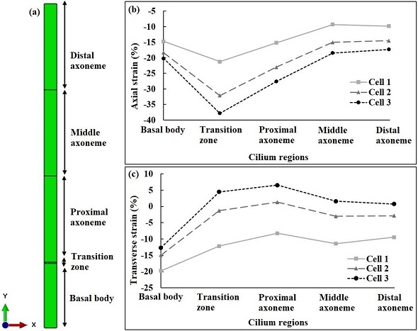
| Figure 3: Cilium strains in RZ chondrocytes at 10% compression. (a) Representation of cilium regions for reporting strains; (b) Depending on the depth of the cell within the RZ the cilium axial (Y-) strains were amplified 2–4X within the cilium transition zone and became more compressive with RZ depth. (b); (c) Cilium strains were amplified 1.25–2X within the basal body region in the transverse (X-) direction and became less compressive with increasing RZ depth and even tensile in more distal regions of the cilium at the deepest location near the PZ (Cell 3). |