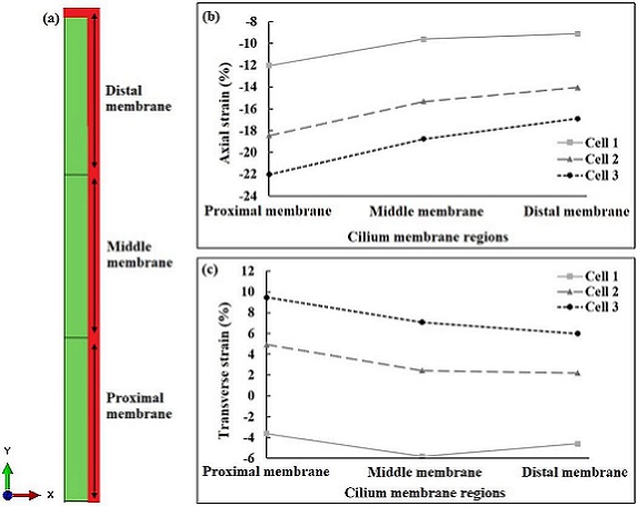
| Figure 4: Cilium membrane strains in RZ chondrocytes at 10% compression. (a) Representation of cilium membrane regions. (b) Axial (Y-) membrane strains were compressive and increased in magnitude with the depth of the cell in the RZ, reaching 2X the applied strain in the cilium proximal membrane region for the cell near the PZ (Cell 3). (c) Transverse to the membrane (X-) strains were tensile for the cell near the PZ (Cell 3) and in the middle of the RZ (Cell 2), but compressive for the cell near the subchondral epiphyseal bone plate. Transverse membrane strain magnitudes decreased distally along the length of the cilium. |